 |
По вопросам ремонта и другим техническим вопросам сюда. Ремонт бытовой и офисной техники.
Подробная информация по лечению, лечебных катушках и оборудованию по телефону +38 067 895 98 24
Или по электронной почте artradiolab@gmail.com
Лечебные катушки - медицинские испытания.
Медицинские испытания Лечебных Катушек доказали уверенное уничтожение ими раковых клеток. Испытания проводились в израильском медицинском центре передовых технологий Шеба. Июль 2017г.
Оригинал отчета на английском языке, кому интересно, скопируйте и вставьте в Гугл Переводчик.
Report on the medical tests of Medical Coils.
Experiment report
Dr. Genady Kostenich
Dr. Mor Oron-Herman
Eng. Katia Kon
Advanced Technology Center
Sheba Medical Center
Goal:
To verify the cytotoxic effect of treatment with the given electric device on cancerous and normal human cells.
Study design:
Human pancreatic carcinoma cells (panc-1) and human fibroblasts (HK3T) were grown in vitro as tissue cultures.
Forty-eight hours before the treatment, the cells were seeded in 48 well tissue culture plate, 25,000 cells/well. Nine central wells were seeded in each plate. Two plates were seeded with fibroblasts, and two plates with pancreatic carcinoma cells. See plate pattern in figure 1.
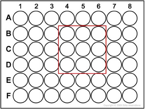
Figure 1: Tissue culture plate seeding pattern.
At the day of treatment, medium was replaced to fresh in order to eliminate LDH residues in medium, coming from spontaneous cell death.
The seeded area of one plate of pancreatic cells was placed between the device transducers for 30 minutes (figure 2), then placed back in the incubator. The same procedure was done with one plate of fibroblasts. The un-treated plates served as control.
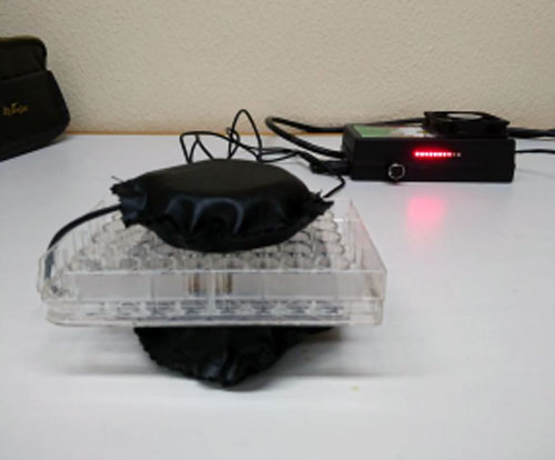
Figure 2: Treatment set up.
LDH-based Cytotoxicity assay:
LDH-based cytotoxicity assay is one of the most commonly used methods for cell death analysis.
Cytotoxic effect of the treatment was measured using CytoTox 96 non- radioactive cytotoxicity assay kit (Promega cat. No. G-1780). The assay quantitatively measures lactate dehydrogenase (LDH), a stable cytosolic enzyme that is released upon cell lysis. Released LDH in culture supernatants is measured with a 30-minute coupled enzymatic assay, which results in conversion of a tetrazolium salt (INT) into a red formazan product. The amount of color formed is proportional to the number of lysed cells.
The general chemical reactions of the CytoTox 96 assay are illustrated in Figure 3.
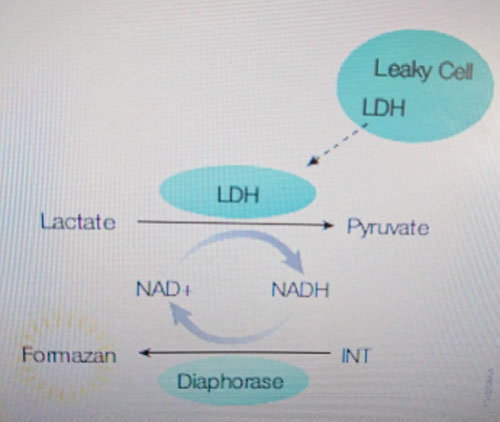
Figure 3: Release of LDH from damaged cells is measured by supplying lactate, NAD+ and INT as substrates in the presence of diaphorase. Generation of a red formazan product is proportional to the amount of LDH released and therefore the number of lysed cells.
Cytotoxicity was measured in each of the 4 plates 6 and 24 hr after treatment.
Percent Cytotoxicity was calculated according to the following formula:
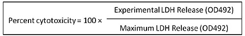
*Experimental LDH release was obtained after subtracting the average values of culture medium background from all values of the experimental wells.
**Maximum LDH was achieved by adding lysis buffer to 3 wells in each plate.
Evans blue staining assay:
Evans blue has proven over the years to be a dependable stain for microscopic determination of cell death. Evans blue is a non-permeating dye. In presence of plasma membrane damage, the dye enters in the cytoplasm and nucleus, thereby staining them blue.
Results:
Cytotoxicity obtained in treated and not treated healthy fibroblasts and pancreatic adenocarcinoma cells is summarized in table 1 and figure 4:
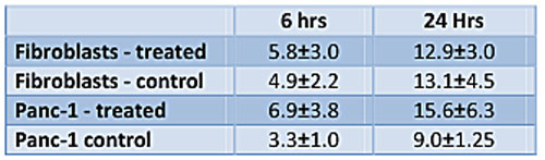
Table 1: Percent cytotoxicity (average±standard deviation) in 4 plates.
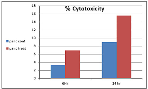
Figure 4: graphic presentation of % cytotoxicity obtained in treated pancreatic cancerous cells compared to non-treated cells 6 and 24 hours after 30 minutes treatment.
The results obtained indicate that there is some selective cytotoxic effect of the treatment on cancerous but not normal human cells. Treated cancerous cells died ~two fold more than non-treated control cells in both time points after treatment. However, the effect is not statistically significant due to standard deviation. No treatment-related cytotoxic effect was found in treated fibroblasts compared to non- treated fibroblasts.
Evans blue staining assay:
At the end of experiment, treated and control pancreatic cancerous cells were stained with Evans Blue dye for microscopic validation of results. The results indicated that most of the cells are alive both in treated and not treated cells. However, the number of stained cells was higher in the treated group compared to untreated control. The effect was heterogeneous: few areas with stained (dead) cells were found in several peripheral wells (zones) in the plate of the treated group (Figure 5).
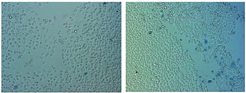
Left - control (not treated) panc 1 cells.
Right - treated panc 1 cells.
Figure 5:
Viability staining of treated and not treated human cancerous pancreatic cells using Evans Blue selective dye. The dye penetrates only into dead (membrane damaged) cells. It can be seen that there are more dead (blue) cells in the treated culture, but still the majority of cells are alive (not stained). Magnification x100. In the viable cells of the treatment group, no visible morphological changes were seen by microscopy under low (x100) and high (x200) magnifications (Fig.5, 6).
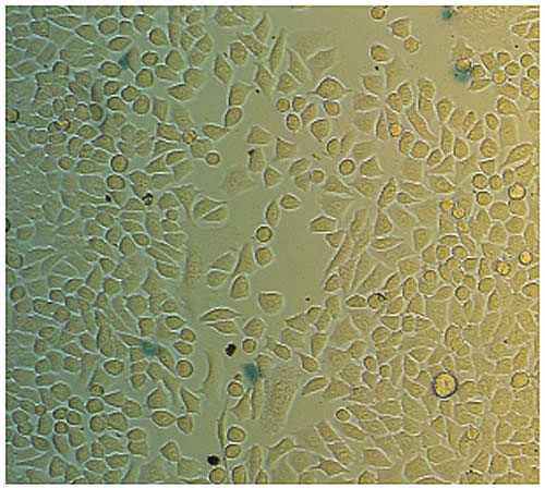
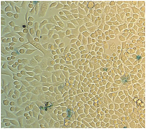
Figure 6, 6a: Human pancreatic cancer cells treated by the device. Magnification x200.
Conclusions:
Slight, however not statistically significant, cancer-selective cytotoxic effect (in comparison with normal fibroblasts) was demonstrated 6 and 24 hrs after 30 minutes treatment using the tested electric device. The effect was heterogeneous: more stained (dead) cells were found in several peripheral wells in the plate of the treatment group. This effect does not seem high enough to destroy tumors in vivo. However, optimization of treatment parameters (e.g. dosage, frequency, geometry of the electric field) and longer follow-up are required in order to reveal the entire potential of the tested treatment.
Выводы о мед испытаниях Лечебных Катушек:
Незначительный, однако статистически недостоверный, селективный (по сравнению с нормальными фибробластами) цитотоксический эффект был продемонстрирован 6 и 24 ч после 30 минут обработки клеток рака поджелудочной железы человека с использованием испытанного электрического устройства.
Эффект был гетерогенным: больше окрашенных (мертвых) клеток было обнаружено в нескольких периферийных зонах и ячейках в плате из группы облучённых клеток.
Этот эффект, возможно, недостаточен для терапии опухолей in vivo. Тем не менее, оптимизация параметров обработки (например, дозы, частоты, геометрии электрического поля) и более длительный период наблюдения необходимы для того, чтобы исследовать весь потенциал тестируемого лечения.
Под заказ можно рассчитать и изготовить любые модификации лечебных катушек.
Статик - прибор для активации воды статическим полем.
РГВП или Optune NovoTTF-100A Лечение вихревыми (TTF) полями.
RGVP or Optune NovoTTF-100A. Treatment by vortex fields.
Печатная плата с разводкой в лайоуте 4.
Генератор для лечебной катушки. Схема. Печатка.
Генератор для лечения статикой. Катушка.
Мишин А. Н. Вихревая динамика
https://www.optune.com
X-FAQ.RU ФОРУМ-1
X-FAQ.RU ФОРУМ-2
ПРИМЕНЕНИЕ КАТУШЕК МИШИНА - ДАНДОРФА.
Как применять лечебные катушки статического резонанса
ЛЕЧЕБНЫЕ КАТУШКИ БЕСПЛАТНАЯ КОНСУЛЬТАЦИЯ.
ЛЕЧЕБНЫЕ КАТУШКИ ИЗГОТОВЛЕНИЕ ПОД ЗАКАЗ.
ВОДА МЕТОДЫ ПОВЫШЕНИЯ ЦЕЛИТЕЛЬНЫХ СВОЙСТВ.
ВОДА КАК ГАРАНТИРОВАНО СТЕРЕТЬ С ВОДЫ ИНФОРМАЦИЮ.
ЛЕЧЕБНЫЕ КАТУШКИ - ПОВЫШЕНИЕ ЭФФЕКТИВНОСТИ.
ЛЕЧЕБНЫЕ КАТУШКИ - УДВОЕНИЕ ЛЕЧЕБНОГО ПОЛЯ.
ЛЕЧЕБНЫЕ КАТУШКИ - УЛУЧШЕННЫЙ ВАРИАНТ.
ЧЕМ ОТЛИЧАЮТСЯ TDA7056, TDA7056A, TDA7056B.
НАШИ РАЗРАБОТКИ.
ЭЛЛАДА-7 АППАРАТ ЭЛЕКТРОПУНКТУРЫ.
ЭЛЛАДА-7 СХЕМА ПЛАТА НАСТРОЙКА.
АППАРАТ ДЛЯ ПОЛУЧЕНИЯ ЖИВОЙ И МЕРТВОЙ ВОДЫ.
АППАРАТ МИКРОМАССАЖА АМ1.
АППАРАТ МИКРОМАССАЖА АМ1 ОТ БОЛЕЗНЕЙ И УШИБОВ.
ЛЕЧЕНИЕ БОЛЕЗНЕЙ АППАРАТАМИ МИКРОМАССАЖА.
С ув. Белецкий А. И. 07.09.2016г. Кубань Краснодар.